


The History of Flow Cytometry and Importance in Research
Flow cytometry is a technique that uses molecules of interest to sort and analyze cells. There are a few existing methods for doing this including acoustics, charge, heavy metal tags, and fluorescence.
The easy favorite among these is “Fluorescence activated cell sorting”, or FACS. FACS adds visualization to cell sorting using dyes and lasers.
The beauty of this is that correlations between molecules are established.
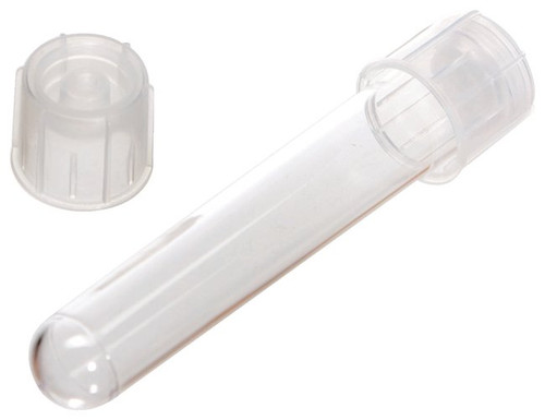
See our Sterile 5mL polystyrene tubes, pictured above
So how does this majestic-sounding technique conjure such amazing results?
The First Step to Isolating Specific Cell Types
Before any meaningful results can be obtained, a “gating strategy” is developed to isolate a specific cell type. These strategies use a molecule to identify one cell type through the process of elimination.
A good example would be the CD markers that human immune cell lineages have.
Although certain subtypes share markers, it’s easy to distinguish one subtype from another by tracking which CD markers are present and which are absent.
The smoke and mirrors here is making sure that you can easily identify markers in your cell samples. A good gating strategy means nothing if you have a poor signal!
With that said, FACS is a very powerful tool when done correctly. Read through a situation next where it has been used to simplify the study question.
FACS Applications in Research
One study looked at the inflammatory marker IFN-α in a high-purity population of dendritic bone marrow cells taken from mice.
They used a surface antigen gating strategy to identify them among similar cell types first by verifying the presence or absence of certain antigens.
Levels of IFN-α were then monitored following treatment with an inflammatory agent and compared between healthy controls and lupus-prone groups and compared to baseline levels.
This protocol allowed purification of a single cell type and quantification of a specific molecule with one straightforward approach.
The feat is the ability to collect a vast amount of data at once. Quick analysis of many molecules is possible, unlike with methods that rely on a human user to identify the targets.
Cell culture analysis has never looked more enchanting! Where to start?!
The takeaway: Methods like this are packed full of elegance, but without the right tools your efforts can go to waste.
FACS Tubes: the Key Unlocking Better Flow Cytometry
FACS tubes are just ordinary clear plastic containers, right? While you can’t make or break your career with them, there's a lot more to them than that.
Let’s talk specifics. These are 5ml containers, shaped like round-bottom cuvettes or conicals.
They are offered as polystyrene or polypropylene plastics. And that’s the key to the proverbial fairy gates here.
If you choose the wrong path, you may lose…
So which is better? Polystyrene is often fragile, and prone to cracks and breaks. However, it offers better clarity and flow cytometry is all about signal detection.
Polypropylene is compatible with more cell types. Fewer kinds stick to these containers and because single cell visualization is the goal, this is a big plus.
That’s it, there’s a balance!
Stellar Brand Features
What are some benefits of Stellar Scientific FACS tubes and accessories?
- Round or pointed bottom.
- Polypropylene or polystyrene.
- Clear plastic.
- Caps for storing samples.
- Strainer caps for reduced workflow.
Our tubes are available in a few select styles and quantities. Unsure what you’ll end up using the most of? Ask us for free samples.
Come see what gifts we have to offer you, traveler…
Considerations for Different Laboratory Settings
Depending on your research scenario and cell types, you might have to coat your collection or sample tubes, or select a specific plastic type.
Polypropylene is the most compatible material available, but coating collection tubes with heparin or fetal calf serum is sometimes necessary.
How much do you need to amplify your signal? If your target is in low abundance and you need amplification, you’ll want a brighter dye and may need a polystyrene tube.
Some of the things you see on the other side of the tube are mere illusions.
The amazing aspect of this method is that you can process up to a dozen different fluorescent targets in a single experiment.
Tips and Tricks for Refining your Protocol
To summarize the key points:
- Strainer caps and coated tubes help to isolate cells for imaging.
- Brighter fluorophores are better for lower abundance targets.
- Choose a fluorescent primary for internal targets.
- Fluorescent secondaries work well for imaging surface molecules.
- Polystyrene is fragile, but it interferes less with signal detection.
Limitations
Some basic limitations include the following:
- The abundance of your target molecule.
- The quality of the sample.
- The number of lasers you are using.
- How well the dyes you chose complement.
There may be other limitations too, but compared to manual sorting methods, this method reigns supreme!
Flow Cytometry Products for Affordable Cost and Quality
The majority of tubes come sterile-packed in bulk bags. We also offer individually wrapped options and non-sterile tubes in larger bulk quantities for a lower cost.
All of our tubes are designed to fit most modern flow cytometry equipment.
See More FACS Tubes and Accessories
Come take a stroll through our full catalog of FACS tubes and accessories. We have different tube materials available for order, and strainer caps to add to any workflow that’s lacking.
The future of flow cytometry analysis is bright. Don’t be left out of the fun!
Not satisfied with the gifts your journey has brought you yet? See the rest of our Flow Cytometry catalog for more cell strainers, reducing adapters, and flow compatible centrifuge tubes to refine the magic of your ideas.
Footnotes:
______________________________________
Bushnell, Tim. “7 Advanced Flow Cytometry Data Analysis Tips For Multi-Color Experiments.” The Cheeky Scientist, expert.cheekyscientist.com/flow-cytometry-data-analysis-tips-for-multi-color-experiments/. Accessed 15 May 2024.
“Flow Cytometry Guide.” Creative Diagnostics, www.creative-diagnostics.com/flow-cytometry-guide... Accessed 15 May 2024.
Herzenberg,LA., Parks, D., Sahaf, B., Perez,O., Roederer, M. (2002) The History and Future of the Fluorescence Activated Cell Sorter and Flow Cytometry: A View from Stanford, Clinical Chemistry, Volume 48, Issue 10, Pages 1819–1827, https://doi.org/10.1093/clinchem/48.10.1819
Liao, X., Makris, M., & Luo, X. M. (2016). Fluorescence-activated Cell Sorting for Purification of Plasmacytoid Dendritic Cells from the Mouse Bone Marrow. Journal of visualized experiments : JoVE, (117), 54641. https://doi.org/10.3791/54641
McKinnon K. M. (2018). Flow Cytometry: An Overview. Current protocols in immunology, 120, 5.1.1–5.1.11. https://doi.org/10.1002/cpim.40
Webber, Jonathan. “FACS Sorting Protocol.” Kennedy Institute of Rheumatology, University of Oxford, www.kennedy.ox.ac.uk/platforms-and-technologies/f... Accessed 15 May 2024.