

Pictures in Pixels - Sensor options and accessories make all the difference in building the best setup for fixed or live-cell digital imaging
Posted by Stellar Staff on 17th Dec 2018
Making microscope images is fundamental to biological and clinical sciences. It’s one thing to see something amazing under a microscope and another to capture that image for further analysis or even just for the wow factor. Instead of shooting rolls of 35-millimeter film, today’s scientists - and those for the past couple decades now - rely on digital methods. Moving this technology ahead depends on companies dedicated to driving this field, and one of the leaders is Motic.
Emerging as a builder of compound microscopes in 1988, Motic started working in the digital world of imaging a decade later.
As the company notes on its website: “This successful transition marked a milestone for the company turning Motic into one of the leading brand names in digital microscopy.” That’s why we carry this company’s products.
To grab a digital image from a microscope, it starts with a digital camera. Various features on a digital camera should be considered.
To give scientists an opportunity to make the most of the best Motic digital camera for a job, we carry a variety of CMOS cameras. Much like consumer digital cameras, resolution makes up a distinguishing feature in a digital cameras for microscopy.
The key is ensuring that a camera provides enough resolution for the expected tasks. With Motic’s CMOS cameras, for example, the Motic Moticam 1 digital camera provides 800 x 600 pixels, and the Moticam 10 provides 3664 x 2748 pixels.
The methods of accessing the images, just like the resolution, keep evolving. So, some digital cameras can now send an image from the stage to a device through a Wi-Fi connection.
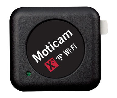
For instance, the Moticam X3 can simultaneously connect with six devices, such as a computer or smartphone. It even allows 4MP streaming over Wi-Fi, which is great for live-cell imaging.
In some cases, scientists need various ways to access images from a digital camera. The Moticam Multi-Output solves that by including USB, HDMI and direct capture to an SD card. That makes this camera a great choice for demonstrations, such as in a teaching lab.
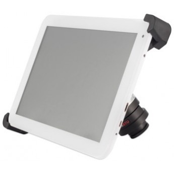
To help a class or lab group watch what’s going on under the scope, some scientists add a tablet to the top of the microscope platform. One option is the Moticam BTU10 , which is a 10-inch Android tablet. It can be used on or off a microscope.
Plus, the Moticam BTU10 can be accessed wirelessly and images can be saved on 16 gigabytes of storage or in various card formats, including SD and others.
The move to digital imaging - powered by a collection of capable platforms and tools - changed what scientists and clinicians could see under a microscope.
Perhaps even more important, digital imaging changes what experts can do with what they see, which ranges from analyzing images in new ways, watching biology in action, sharing science around the world or teaching tomorrow’s experts.
Then, those images or movies can be stored and retrieved across the years.
Those capabilities add analytical opportunities to basic and clinical research, some of which cannot be accomplished without digital imaging.