

Get Clear and Reliable Results with Our Gel Documentation Systems
21st Aug 2025
In molecular biology and biochemistry, clear and reproducible documentation of gel electrophoresis results is essential. From DNA fragment analysis to protein quantification, researchers depend on high-quality imaging systems to visualize and analyze their gels with precision. Reliable data capture not only saves time but also ensures experimental reproducibility and data integrity.
That’s where a high-performance gel documentation system comes into play. These systems are indispensable tools in any laboratory that routinely uses agarose or polyacrylamide gel electrophoresis. Whether you're conducting routine diagnostics or publishing high-impact research, clear and reliable imaging is key to success.
What Is a Gel Documentation System?
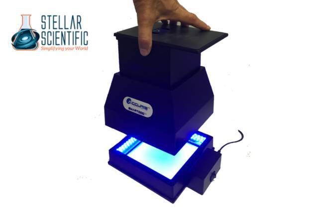
A gel documentation system, often referred to as a gel doc, is an imaging setup used to capture high-resolution images of DNA, RNA, or protein gels stained with various dyes. These systems combine sensitive cameras, light sources, filters, and analysis software into a single unit that makes it easy to document and quantify bands on electrophoresis gels.
They replace outdated, manual methods such as using Polaroid cameras or film, offering instant image capture, enhanced clarity, and digital file storage. With a modern gel doc system, users can quickly adjust exposure, analyze band intensity, and export data for further use.
Key Features of Our Gel Documentation Systems
1. High-Resolution Imaging
One of the most critical aspects of a gel documentation system is its ability to capture fine detail. Our systems use high-resolution CCD or CMOS cameras designed specifically for scientific imaging. These cameras provide superior sensitivity and dynamic range, making it easier to detect faint bands while avoiding overexposure of brighter areas.
Whether you’re analyzing nucleic acids stained with ethidium bromide or proteins labeled with Coomassie blue, image clarity is consistent and precise.
2. Versatile Light Sources
Different applications require different types of illumination. Our gel documentation systems offer a variety of lighting options, including:
- UV transilluminators for detecting ethidium bromide or SYBR stains
- Blue light LEDs for safe imaging of fluorescent dyes like SYBR Safe
- White light conversion screens for protein gels stained with silver or Coomassie
These options provide flexibility for researchers using various gel stains and minimize the risk of photodamage to sensitive samples.
3. Easy-to-Use Interface and Software Integration
A user-friendly interface is essential in fast-paced lab environments. Our gel documentation systems feature intuitive touchscreens or software controls that streamline the imaging process. Real-time preview, auto-focus, and adjustable exposure settings enable quick optimization of image capture.
Paired with advanced analysis software, users can:
- Measure band intensity
- Calculate molecular weights using ladders
- Normalize signals for quantitative analysis
- Annotate and export results in multiple formats
This streamlined workflow saves time and enhances the accuracy of your gel analysis.
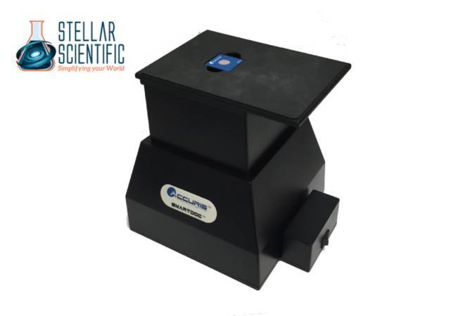
4. Compact and Ergonomic Design
Bench space is often limited, especially in smaller labs. Our gel doc systems are compact and ergonomically designed to fit comfortably on standard lab benches. The enclosed imaging chambers also ensure user safety when working with UV light, with interlock systems that disable UV exposure when the door is open.
5. Reproducibility and Data Integrity
Digital gel documentation ensures that imaging results can be saved, retrieved, and compared across experiments. With automated settings and programmable templates, our systems support standardized workflows that reduce variability between runs. This helps meet data reproducibility standards and supports high-quality research outputs.
6. Low Maintenance and Long-Term Reliability
Our gel documentation systems are built to perform reliably over time with minimal maintenance. LED lighting components are long-lasting and energy-efficient, while the sealed imaging chambers help reduce contamination and equipment wear. With proper care, these systems continue delivering consistent performance even in high-use environments.
Common Applications of Gel Documentation Systems
DNA and RNA Analysis
After running a gel electrophoresis, visualizing DNA or RNA bands stained with intercalating dyes is a standard practice in molecular biology. Gel documentation instruments enable researchers to detect and measure fragment sizes with precision. This is particularly important for verifying PCR results, performing restriction digests, or checking cloning success.
Protein Gel Imaging
Whether using SDS-PAGE or native PAGE, researchers need to document protein separation and staining. Our systems accommodate white light illumination for visualizing Coomassie or silver-stained gels. Band intensity analysis also allows for semi-quantitative comparison of protein expression levels.
CRISPR and Genotyping Verification
Gene editing and genotyping workflows rely heavily on accurate fragment analysis. Researchers can use gel docs to confirm indels or successful gene insertions by comparing expected and observed band patterns. High-resolution imaging ensures that even subtle differences are clearly visible.
Education and Training
In academic settings, clear gel images are valuable for teaching students fundamental lab techniques. Instructors can capture and project gel results for class demonstrations, improving understanding and engagement during lab sessions.
Choosing the Right Gel Documentation System
When selecting a gel doc system for your lab, consider the following:
- Camera resolution and sensitivity: For detecting faint bands, higher resolution and greater sensitivity are essential.
- Compatibility with stains: Make sure the system supports the types of dyes you commonly use.
- Software features: Look for systems with easy data export, band analysis, and ladder comparison tools.
- Size and safety: Compact systems with UV protection and easy-clean surfaces are ideal for shared labs.
- Connectivity: Many systems now offer USB, Ethernet, or Wi-Fi connections for data transfer and remote access.
Choosing the right system ensures you get the best possible results from your electrophoresis workflows while optimizing time and space in the lab.
Tips for Better Gel Imaging
To make the most of your gel documentation system:
- Use clean, well-prepared gels to reduce background noise.
- Avoid overloading wells, which can cause band smearing.
- Optimize exposure settings to prevent under- or overexposed bands.
- Always use proper safety equipment when working with UV light.
- Save original image files before editing to maintain raw data integrity.
These simple practices help ensure clear, reliable documentation for every experiment.
Why Labs Trust Our Gel Documentation Systems
Laboratories across academia, clinical diagnostics, and biotech choose our lab gel documentation system for their ease of use, image quality, and long-term reliability. Whether you're imaging DNA, RNA, or proteins, our systems provide the flexibility and performance needed to support a wide range of molecular biology applications.
Reliable imaging isn’t just about convenience; it’s about data quality, reproducibility, and research integrity. With our systems, users can focus more on science and less on troubleshooting equipment.
About Stellar Scientific
Stellar Scientific is a trusted provider of high-quality laboratory equipment and consumables. We serve research institutions, universities, clinical labs, and biotechnology companies with products that support a broad range of scientific applications. Our offerings include gel documentation systems, centrifuges, incubators, pipette tips, gloves, cleaning agents, and much more. At Stellar Scientific, we are committed to delivering performance, reliability, and value through every product we supply.